Sagittal view of sheep brain – Step into the fascinating realm of the sheep brain’s sagittal view, where intricate structures and neural pathways unfold before our eyes. This view, like a window into the brain’s inner workings, grants us a unique perspective on the organ that governs our thoughts, emotions, and actions.
The sagittal view, slicing through the brain from front to back, reveals a landscape of anatomical marvels. The cerebrum, the brain’s command center, dominates the scene, while the cerebellum, responsible for coordination and balance, nestles beneath it. The brainstem, the vital connection between the brain and spinal cord, emerges from the depths, carrying essential life-sustaining signals.
Overview of Sagittal View of Sheep Brain

The sagittal view of the sheep brain is an anatomical perspective that provides a midline cross-section of the brain, dividing it into left and right hemispheres. This view allows for a comprehensive examination of the brain’s internal structures and their spatial relationships.
In the sagittal view, the brain appears as an elongated, oval-shaped structure. The cerebrum, the largest part of the brain, is located at the front and occupies the majority of the cranial cavity. Behind the cerebrum lies the cerebellum, which is responsible for coordinating movement and balance.
The brainstem, which connects the cerebrum and cerebellum to the spinal cord, is situated at the base of the brain.
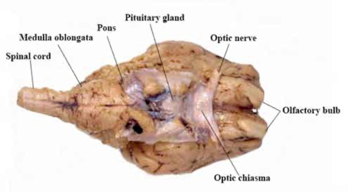
The sagittal view of the sheep brain provides a detailed look at its internal structures. The cerebral hemispheres, cerebellum, and brainstem are all clearly visible. It’s interesting to note that the sheep brain has a relatively small olfactory bulb compared to humans, which may be related to their reduced reliance on smell.
Just like humans, sheep can also suffer from alergia a la luz uv led . This condition, which causes an allergic reaction to ultraviolet light, can lead to skin irritation, itching, and swelling. Back to the sagittal view of the sheep brain, the hypothalamus and pituitary gland are also visible, which are important for regulating various bodily functions.
Internal Structures
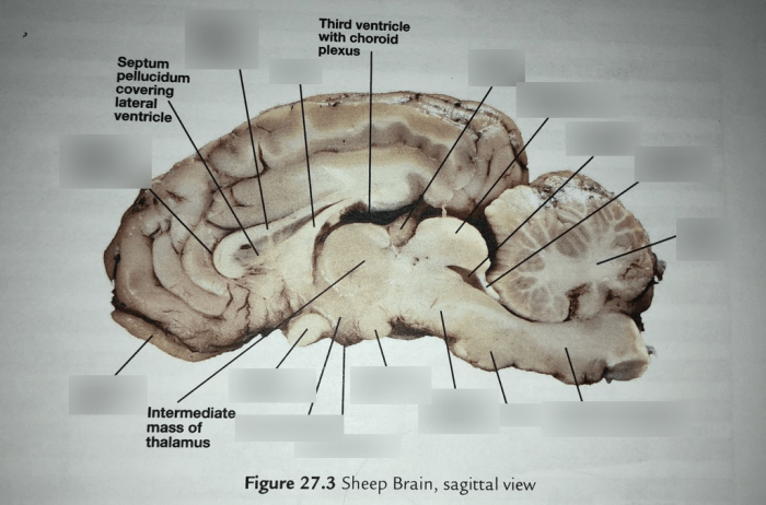
The sagittal view reveals various internal structures within the brain. These include:
- Cerebral Hemispheres:The two hemispheres of the cerebrum are separated by a deep groove called the longitudinal fissure. Each hemisphere is responsible for different cognitive functions, such as language, memory, and reasoning.
- Corpus Callosum:A thick band of nerve fibers that connects the two cerebral hemispheres, facilitating communication between them.
- Thalamus:A relay center for sensory information, which directs sensory signals to the appropriate areas of the cerebrum for processing.
- Hypothalamus:A small region below the thalamus that regulates body temperature, hunger, thirst, and other homeostatic functions.
- Pituitary Gland:A small gland attached to the hypothalamus that secretes hormones essential for growth, development, and metabolism.
- Cerebellum:A structure located at the back of the brain that coordinates movement and balance.
- Brainstem:The brainstem connects the cerebrum and cerebellum to the spinal cord and is responsible for vital functions such as breathing, heart rate, and blood pressure.
Anatomical Structures Visible in Sagittal View

The sagittal view of the sheep brain provides a comprehensive perspective of its internal structures. This view allows us to examine the brain’s intricate anatomy, including its major components and their spatial relationships.
The most prominent structure visible in the sagittal view is the cerebrum, the largest part of the brain. It occupies the anterior portion of the cranial cavity and is divided into two hemispheres, separated by the falx cerebri. The cerebrum is responsible for higher-order cognitive functions such as consciousness, perception, and voluntary movement.
Posterior to the cerebrum lies the cerebellum, a smaller but equally important structure. The cerebellum plays a crucial role in coordinating movement, balance, and posture.
Beneath the cerebrum and cerebellum, the brainstemconnects the brain to the spinal cord. The brainstem consists of the midbrain, pons, and medulla oblongata, each of which serves specific functions related to sensory and motor control, as well as vital life-sustaining processes such as breathing and heart rate regulation.
Within the brain, there are also several fluid-filled cavities called ventricles. The ventricles produce and circulate cerebrospinal fluid, which provides nutrients and protection to the brain tissue.
The sagittal view also reveals the major blood vesselsthat supply the brain. The internal carotid arteries and vertebral arteries bring oxygenated blood to the brain, while the internal jugular veins drain deoxygenated blood away.
Functional Significance of Sagittal View

The sagittal view offers a unique perspective of the brain, providing valuable insights into its intricate structure and functionality. It allows researchers and clinicians to visualize the brain’s midline structures, neural pathways, and connections, aiding in the understanding of brain function and dysfunction.
This view is particularly useful in studying the interconnections between different brain regions. By examining the sagittal section, researchers can trace the course of nerve fibers as they connect various parts of the brain, forming complex neural circuits responsible for cognitive processes, sensory perception, and motor control.
Applications in Medical Imaging and Research, Sagittal view of sheep brain
The sagittal view has significant applications in medical imaging and research. It is commonly employed in:
- Magnetic resonance imaging (MRI):MRI scans provide detailed sagittal images of the brain, enabling the visualization of brain structures, lesions, and abnormalities.
- Computed tomography (CT):CT scans offer high-resolution sagittal images of the brain, useful for diagnosing skull fractures, tumors, and other structural abnormalities.
- Neurosurgical planning:The sagittal view assists neurosurgeons in planning surgical procedures by providing a clear understanding of the brain’s anatomy and the location of critical structures.
- Neurological research:Researchers use the sagittal view to study brain development, connectivity, and function in both healthy and diseased individuals.
Clinical Applications of Sagittal View
The sagittal view of the sheep brain is a valuable tool in clinical settings, providing insights into brain structure and function. It plays a crucial role in diagnosing and treating various brain disorders, enabling medical professionals to make informed decisions and develop effective treatment plans.
Diagnostic Applications
- Tumors and Lesions:The sagittal view allows visualization of tumors, cysts, and other lesions within the brain. By examining the size, shape, and location of these abnormalities, clinicians can determine their nature and plan appropriate interventions.
- Hydrocephalus:The sagittal view helps detect hydrocephalus, a condition characterized by excessive cerebrospinal fluid accumulation within the ventricles. By assessing the size and shape of the ventricles, clinicians can diagnose and monitor hydrocephalus, guiding treatment decisions.
- Infections:The sagittal view can reveal signs of brain infections, such as abscesses or encephalitis. By observing changes in brain tissue density and the presence of fluid collections, clinicians can identify and localize infections, facilitating prompt treatment.
Treatment Planning
- Neurosurgery:The sagittal view provides essential anatomical information for neurosurgical procedures. It helps surgeons plan surgical approaches, identify critical structures, and minimize risks during interventions.
- Radiation Therapy:The sagittal view aids in planning radiation therapy for brain tumors. By accurately defining the tumor’s location and extent, clinicians can optimize radiation delivery, maximizing treatment effectiveness while minimizing damage to healthy tissue.
Comparison with Other Brain Views

The sagittal view provides a unique perspective of the brain compared to other commonly used views, such as the axial and coronal views. Each view offers distinct advantages and limitations, contributing to a comprehensive understanding of brain anatomy and function.
Axial View
The axial view, also known as the transverse view, is obtained by cutting the brain horizontally, parallel to the base of the skull. It provides cross-sectional images of the brain, allowing for the visualization of structures at different levels along the anterior-posterior axis.
The axial view is particularly useful for identifying the relationship between different brain regions and for studying the internal structures of the brain, such as the ventricles, basal ganglia, and thalamus.
Coronal View
The coronal view, also known as the frontal view, is obtained by cutting the brain vertically, perpendicular to the base of the skull. It provides images of the brain from the front to the back, allowing for the visualization of structures along the medial-lateral axis.
The coronal view is particularly useful for studying the relationship between different brain regions within a hemisphere and for identifying the location of sulci and gyri.
Complementary Nature of Different Views
The sagittal, axial, and coronal views complement each other in providing a comprehensive understanding of brain anatomy and function. By combining information from different views, it is possible to obtain a more complete picture of the brain’s structure and organization.
For example, the sagittal view can provide information about the overall shape and size of the brain, while the axial and coronal views can provide more detailed information about the internal structures and their relationships to each other.
FAQ Insights: Sagittal View Of Sheep Brain
What is the significance of the sagittal view of the sheep brain?
The sagittal view provides a detailed cross-sectional view of the brain, allowing for the visualization of anatomical structures, their relationships, and neural connections.
How is the sagittal view used in clinical settings?
The sagittal view is employed in diagnosing and treating brain disorders, such as tumors, hemorrhages, and developmental abnormalities, by providing a comprehensive view of the brain’s internal structures.
What are the advantages of using the sagittal view compared to other brain views?
The sagittal view offers a unique perspective on the brain’s midline structures, including the corpus callosum, brainstem, and ventricular system, which are not as clearly visible in other views.